Wednesday, November 26, 2014

BURSITIS AND TENDINITIS AROUND HIP


TROCHANTERIC BURSITIS


Pain over the lateral aspect of the hip and thigh may be due to local trauma or overuse, resulting in inflammation of the trochanteric bursa which lies deep to the tensor fascia lata. There is local tenderness and sometimes crepitus on flexing and extending the hip. Swelling is unusual but post-traumatic bleeding can produce a bursal haematoma.


X-ray may show evidence of a previous fracture or a protruding metal implant or trochanteric wires dating from some former operation. There may also be calcification or shadows suggesting swelling of the soft tissue. It is important to exclude underlying disorders such as Gout, Rheumatoid Arthritis and infections (including Tuberculosis)           

Other causes of pain and tenderness over the greater trochanter are stress fracture (in athletes and elderly patients), slipped epiphysis (in adolescents) and bone infections (in children). The common cause of misdiagnosis is referred pain from the lumbar spine.
The usual treatment is rest, Physiotherapy, NSAIDs, and injection of local anaesthetic and corticosteriod (provided infection is excluded). If a haematoma is present should be drained.

In Physiotherapy, cold compression, ultrasonic therapy and TENS can help reduce pain and swelling.


GLUTEUS MEDIUS TENDINITIES

Acute tendinitis may cause pain and localized tenderness just behind the greater trochanter. This is seen particularly in dancers and athletes. The clinic finding and X-ray features are similar to those of trocanteric bursitis, andthe deferential diagnosis is the same. Treatment protocol is also almost the same.


ADDUCTOR LONGUS STRAIN OR TENDINITIS

This overuse injury is often seen in footballers and athletes. The patients complains of pain in the groin and tenderness can be localized to the adductor longus origin, close to pubis. Swelling below this site may signify an adductor longus tear. 
Acute strains are treated with rest and heat., while chronic strains may need prolonged PHYSIOTHERAPY.


ILIOPSOAS BURSITIS

Pain in the groin and anterior thigh may be due to an iliopsoas bursitis. The site of tenderness is difficult to define and there be guarding of the mussels overlying the lesser trochanter. Hip movements are sometimes restricted; indeed, the condition may arise from synovitis of the hip joint since there is often potential communication between the bursa and joint.

The most typical feature is a sharp pain on adduction and internal rotation of the hip. Pain is also elicited by testing psoas contraction against resistance.
The differential diagnosis of anterior hip pain includes lymphadenopathy, hernia, a psoas abscess, fracture of the lesser trochanter, slipped epiphysis, local infection and arthritis.


SNAPPING HIP


Snapping hip is a disorder in which the patient  (usually women) complains of hip ‘jumping out of place, or catching’, during walking. The snapping is caused by a thickened band in the gluteus maximus aponeurosis flipping over the greater trochanter. In the swing phase of walking the band moves anteriorly; then, in the stance phase, as the gluteus maximus contracts and pull the hip in extension, the band slips back across the trochanter causing an audible snap. This is usually painless but can be quite distressing, especially if the hip gives way. Sometimes there is tenderness around the hip, and it may be possible to reproduce the peculiar sensation by flexing and extending the hip while the patient contracts the abductors.

Treatment of the snapping tendon is usually unnecessary, the patient merely needs an explanation and reassurance. Occasionally, though if discomfort is marked, the band can be either divided or lengthened by Z- plasty.


referances:
solomon
mendmeshop pics


for any assistance for pain around hip,
contact PAIN FREE PHYSIOTHERAPY CLINIC
31 A, DDA Flats, Pocket 2, Behind Sec 6
Dwarka, New Delhi 110075


Friday, July 11, 2014

Ankle sprain or twisted ankle

A sprained ankle or twisted ankle is a common cause of ankle pain.  
More common is an inversion sprain (or lateral collateral ligament sprain) where the ankle turns over so the sole of the foot faces inwards, damaging the ligaments on the outside of the ankle.

A medial ligament sprain is rare but can occur particularly with a fracture. This happens when the ankle rolls the other way, so the sole of the foot faces outwards, damaging the ligaments on the inside of the ankle. 

The lateral collateral ligament comprises of 3 bands connecting the fibula anteriorly and posteriorly with the talus and with a 3rd band on the sides with the calcaneum. Severe the injury more the no. of bands injured.

In addition to the ligament damage there may also be damage to tendons, bone and other joint tissues, that is why it is important to get a professional to diagnose your ankle sprain.


                                          Grades of Severity for Sprained Ankles:

Sprained ankles, as with all ligaments sprains, are divided into grades.

Grade 1 sprain:

  • Some stretching or perhaps minor tearing of the lateral ankle ligaments.
  • Little or no joint instability. 
  • Mild pain.
  • There may be mild swelling around the bone on the outside of the ankle.
  • Some joint stiffness or difficulty walking or running.


Grade 2 sprain:

  • Moderate tearing of the ligament fibres.
  • Some instability of the joint.
  • Moderate to severe pain and difficulty walking.
  • Swelling and stiffness in the ankle joint.
  • Minor bruising may be evident.


Grade 3 sprain:

  • Total rupture of a ligament.
  • Gross instability of the joint.
  • Severe pain initially followed later by no pain.
  • Severe swelling.


                                                         Treatment of a Sprained Ankle

Immediate First Aid for a sprained ankle:
Aim to reduce the swelling by PRICE as soon as possible.

P is for PROTECTION. The injured ankle should be protected with utmost care to prevent any further injury.

R is for REST. It is important to rest the injury to reduce pain and prevent further damage. Use crutches it necessary. Many therapists advocate partial weight bearing as soon as pain will allow. This is thought to accelerate rehabilitation.

I is for ICE or cold therapy. Applying ice and compression can ease the pain, reduce swelling, reduce bleeding (initially) and encourage blood flow (when used later). Apply an ice pack or similar immediately following injury for 15 minutes. Repeat this every 2 hours.

C is for COMPRESSION - This reduces bleeding and helps reduce swelling.

E is for ELEVATION - Uses of gravity to reduce bleeding and swelling by allowing fluids to move away from the site of injury.


Further treatment

In cases of severely injured ankle (grade 2 and 3) cast can be applied for 3-6 week as per the orthopaedics decision. At times surgical intervention may be needed.

Following the initial painful stage in grade 1 treated with PRICE and after removal of cast in severe cases, physiotherapy treatment can help the ankle return to normal as soon as possible.

Physiotherapeutic modalities like LASER therapy, Ultrasonic therapy and TENS can help reduce pain further and promote healing. Hot fomentation can increase the circulation of the area which further promote healing and increase the pliability of the tissues. Wax bath instead of hot fermentation can be used in cases where cast was applied. 

To strengthen the soft tissues around the ankle start with gentle isometric (tension building without movement) exercises, followed by range of motion exercises within the painfree limits. As for e.g.. writing the alphabets A-Z with the great toe on the floor, heal raises in sitting etc. Approach your physiotherapist to learn more.

Strength can be further developed though resisted exercises with the help of therabands.

The techniques like cross frictional massage and ligament stretching can be implemented to prevent adhession formation and to maintain the flexibility of the ligaments. This helps to reduce the chances of recurrent ankle sprain problems.

The calf muscles often tighten up to protect the joint following a sprained ankle, and so gently stretching the calf muscles can also help to maintain movement at the joint.

A wobble balance board is an important part of rehabilitation of ankle sprains to improve the proprioception (joint sense) which generally gets hampered after any injury.

Contact Pain Free Physiotherapy Clinic for Physiotherapy treatment of ankle sprain.

Dr. Roshan Jha (PT)
Sr. Physiotherapist 
Pain Free Physiotherapy Clinic
31 A, DDA Flats, Pocket 2, Sector 6, Dwarka
Ph. No. 8800299652

Tuesday, July 8, 2014

KNEE JOINT AND ASSOCIATED CONDITIONS

                    KNEE JOINT

The knee is a hinge-joint formed between the tibia (lower bone) and femur (upper bone), also called Tibiofemoral joint, which is dived into medial and lateral compartment. The patella glides over the front of femoral condyles to form a patellofemoral joint. In addition, there is superior tibiofibular joint between the tibia and the head of fibula(outer leg bone).
                                                                                                                                                                         The knee joint is particularly susceptible to traumatic injury because it depends more on the ligaments due to less musculature around it for its strength and stability.  


The space between the tibia and femur is partially filled by two menisci (inner and outer sides) that are attached to add congruency. They aid in lubrication and nutrition of the joint and act as shock absorbers.

The functions of different ligaments of the knee are:
LIGAMENT

FUNCTION
Medial Collateral (inner side ligament)

Prevents medial (inner side) opening up

Lateral Collateral (outer side ligament)
Prevents lateral (outer side) opening up

Anterior Cruciate (within joint on the front side)
Prevents anterior (front) translation of the tibia on the femur

Posterior Cruciate (within joint on the back side)
Prevents posterior (back) translation of the tibia on the femur.
It must also be remembered that the lumbar spine (lower back), hip, and ankle may refer pain to the knee, and these joints must be assessed if it appears that joints other than the knee may be involved.


CLINICAL SIGNIFICANCE:
§              
     Chondromalacia patella (also called patellofemoral syndrome): Irritation of the cartilage on the underside of the kneecap (patella), causing knee pain. This is a common cause of knee pain in young people.

§        Knee osteoarthritis: Osteoarthritis is the most common form of arthritis, and often affects the knees. Caused by aging and wear and tear of cartilage, osteoarthritis symptoms may include knee pain, stiffness, and swelling.

Knee effusion: Fluid buildup inside the knee, usually from inflammation. Any form of arthritis or injury may cause a knee effusion.

§      Meniscal tear: Damage to a meniscus, the cartilage that cushions the knee, often occurs with twisting the knee. Large tears may cause the knee to lock.

§       ACL (anterior cruciate ligament) strain or tear: The ACL is responsible for a large part of the knee’s stability. An ACL tear often leads to the knee “giving out,” and may require surgical repair.


§   PCL (posterior cruciate ligament) strain or tear: PCL tears can cause pain, swelling, and knee instability. These injuries are less common than ACL tears, and physical therapy (rather than surgery) is usually the best option.

§      MCL (medial collateral ligament) strain or tear: This injury may cause pain and possible instability to the inner side of the knee.

Patellar subluxation: The kneecap slides abnormally or dislocates along the thigh bone during activity. Knee pain around the kneecap results.

§     Patellar tendonitis: Inflammation of the tendon connecting the kneecap (patella) to the shin bone. This occurs mostly in athletes from repeated jumping.


PAIN CHARACTERISTICS AND THE POSSIBLE LESION
1
Sharp catching pain
Mechanical defect (trauma)
2
Aching type pain with morning stiffness which eases with movements but gets worst with exertion
Degenerative changes
3
Night pain
Degenerative changes, lateral cystic cartilage, Meniscal tear
4
Pain on weight bearing
Mechanical or arthritic lesion
5
Pain while climbing up or down the steps or rising from chair
Patellofemoral origin
6
Pain on twisting
Meniscal origin
7
Pain over bony eminence below knee in young adults
Osgood Schlatter’s disease


                                           Physiotherapy & Exercise


We all know that your physiotherapist is an expert in the prescription of exercise appropriate to you and your injury or fitness level. As a part of their physiotherapy training, your physiotherapist not only is educated in injury diagnosis but also in exercise physiology or the science of exercise. This enables your physiotherapist to not only assess and diagnose your injury but also to prescribe injury, fitness or age-appropriate exercises targeted to you at that point in time.


What Exercises Should You Do?

It is important that your exercises should not be painful. While some personal trainers believe that the more painful the better, this is not the best for your body or injury. 
In fact, research does inform us that pain inhibits muscles from performing to their optimum. This argues the case that painful exercise is actually counter-productive.
You'll find that your physiotherapist will thoroughly examine you and prescribe a series of exercises suitable for you in quantities that will not injure you further. Please seek an exercise expert, such as your physiotherapist, when you are planning your rehabilitation.


What Happens When You Stop Exercises?

Without some simple exercises, we know that certain muscles can become weak. When these supporting muscles are weak, your injured structures are inadequately supported and predispose you to lingering symptoms or further injury. You can also over-activate adjacent muscles that may lead to further injury.

It is also important to understand that even if you are "in good shape," you may have weak localised or stability muscles.
When you have an injury, you should perform specific exercises that specifically strengthen the muscles around your injury and the adjacent joints. Your physiotherapist will assess your muscle function and prescribe the right exercises specific for your needs.

The exercises prescribed will usually be relatively simple, and do not require any special weights equipment, and can be performed safely at home.

Would You Stop Your Daily Prescribed Drugs?

When your physiotherapist prescribes your individualised dose or exercises, they are using their professional expertise to optimise your exercise dose. 

Would you just stop taking your regular blood pressure medication because you were too busy or didn't think it was working?  I would hope not!

Exercise when prescribed by an expert such as your physiotherapist should be treated as your mandatory dose as prescribed by your physiotherapist. Just like when you don;t take your blood pressure medication, you can't expect the drugs to work of you don't take it as prescribed by your health professional!
So, next time you skip your "exercise dose" just remember that you are not putting your health first.


If you have any questions, please contact PAIN FREE PHYSIOTHERAPY CLINIC.


Dr. AMITA VARSHNEY (PT)
BPT, DCPT, Mulligan Practitioner
PAIN FREE PHYSIOTHERAPY CLINIC
31-A, DDA Flats,Pkt II, Sector 6, Dwarka, New Delhi 110075
Ph. no. - 011-45020554, +918800299652


Tuesday, June 24, 2014

BACK PAIN (All you need to know)

Back pain is one of the most common medical problems, affecting 9 out of 10 people at some point during their lives. Back pain can range from a dull, constant ache to a sudden, sharp pain. This pain or discomfort can happen anywhere in your back, the most common area affected is your low back. This is because the low back supports most of your body's weight. Acute back pain comes on suddenly and usually lasts from a few days to a few weeks. Back pain is called chronic if it lasts for more than three months.


Acute Low Back Ache

Acute low back pain is most often caused by a sudden injury to the muscles and ligaments supporting the back. The pain may be caused by muscle spasms or a strain or tear in the muscles and ligaments.

Causes of sudden low back pain include:

1. Compression fractures to the spine from osteoporosis
2. Cancer involving the spine
3. Fracture of the spinal cord
4. Muscle spasm (very tense muscles)
5. Ruptured or herniated disk
6. Sciatica (numbness/ tingling running down the leg)
7. Spinal stenosis (narrowing of the spinal canal)
8. Spine curvatures (like scoliosis or kyphosis), which may be inherited and seen in children
9. Strain or tears to the muscles or ligaments supporting the back


chronic low back ache

It may result from arthritic (wear and tear  that occurs over the year) changes, which may be due to -

1. Heavy use from work or sports
2. Past injuries and fractures
4. Surgical intervention of the spine in the past
5. Discal changes over the years resulting from herniation of disc which once upon a time was an acute problem

other causes can be..

6. long standing cases of scoliosis and kyphosis
7. Medical problems, such as fibromyalgia, rheumatoid arthritis, and psoriatic arthritis


Symptoms

Low back pain can vary widely. The pain may be mild, or it can be so severe that you are unable to move.A variety of symptoms depending upon the cause can appear in the back, buttock region, thigh and at times till toes. You may have a tingling or burning sensation, a dull achy feeling, or sharp pain, weakness in your legs or feet.


You are at greater risk for low back pain if you:

1. Are over age 30
2. Are overweight
3. are pregnant
4. Do not exercise
5. Feel stressed or depressed
6. Have a job in which you have to do a lot of heavy lifting, bending and twisting, or that involves whole body vibration (such as truck driving or using a sandblaster)
7. Smoke


                                                      BACK CARE AT HOME

A common myth about back pain is that you need to rest and avoid activity for a long time. In fact, doctors do not recommend bed rest. If you have no sign of a serious cause for your back pain (such as loss of bowel or bladder control, weakness, weight loss, or fever), stay as active as possible.


Here are tips for how to handle back pain and activity:

1. Stop normal physical activity for only the first few days. This helps calm your symptoms and reduce swelling (inflammation) in the area of the pain.
2. Apply heat or ice to the painful area. Use ice for the first 48 to 72 hours, then use heat.
3. Take over-the-counter pain relievers such as ibuprofen or acetaminophen.
4. Sleep in a curled-up, fetal position with a pillow between your legs. If you usually sleep on your back, place a pillow or rolled towel under your knees to relieve pressure.
5. Do not do activities that involve heavy lifting or twisting of your back for the first 6 weeks after the pain.
6. Do not exercise in the days right after the pain begins. After 2 to 3 weeks, slowly begin to exercise again.

A physiotherapist can teach you which exercises are right for you.


A complete exercise program should include aerobic activity (such as walking, swimming, or riding a stationary bicycle), as well as stretching and strength training. Follow the instructions of your physiotherapist.

Begin with light cardiovascular training. Walking, riding a stationary bicycle, and swimming are great examples. These types of aerobic activities can help improve blood flow to your back and promote healing. They also strengthen muscles in your stomach and back.

Stretching and strengthening exercises are important in the long run. Keep in mind that starting these exercises too soon after an injury can make your pain worse. Strengthening your abdominal muscles can ease the stress on your back. A physiotherapist can help you determine when to begin stretching and strengthening exercises and how to do them.

Avoid these exercises during recovery, unless your doctor or physiotherapist say it is okay:

Jogging
Contact sports
Racquet sports
Golf
Dancing
Weight lifting
Leg lifts when lying on your stomach
Sit-ups

TAKING MEASURES TO PREVENT FUTURE BACK PAIN

To prevent back pain, learn to lift and bend properly. Follow these tips:

If an object is too heavy or awkward, get help.
Spread your feet apart to give you a wide base of support.
Stand as close as possible to the object you are lifting.
Bend at your knees, not at your waist.
Tighten your stomach muscles as you lift or lower the object.
Hold the object as close to your body as you can.
Lift using your leg muscles.
As you stand up while holding the object, do not bend forward.
Do not twist while you are bending to reach for the object, lifting it up, or carrying it.

Other measures to prevent back pain include:

Avoid standing for long periods. If you must stand for your work, place a stool by your feet. Alternate resting each foot on the stool.
Do not wear high heels. Wear shoes that have cushioned soles when walking.
When sitting, especially if using a computer, make sure that your chair has a straight back with an adjustable seat and back, armrests, and a swivel seat.
Use a stool under your feet while sitting so that your knees are higher than your hips.
Place a small pillow or rolled towel behind your lower back while sitting or driving for long periods.
If you drive long distance, stop and walk around every hour. Do not lift heavy objects just after a long ride.
Quit smoking.
Lose weight.
Do exercises to strengthen your abdominal muscles. This will strengthen your core to decrease the risk of further injuries.
Learn to relax. Try methods such as yoga, tai chi, or massage.


(courtesy-NIH-US)


If the symptoms doesn't improve contact PAIN FREE PHYSIOTHERAPY CLINIC.

Dr. Roshan Jha(PT)
Sr. Physiotherapist
Pain Free Physiotherapy Clinic

Saturday, June 21, 2014

Role of Physiotherapy in Elderly People (Geriatric Population)

                                                   
Physiotherapy keeps elderly physically active

Regular physical activity has been shown to have important beneficial effects on physical and mental well being across all age groups. There are very few medical conditions that regular exercise doesn't help to prevent, reduce the risk of developing, or improve symptoms.
Physical activity in older adults can benefit from participation in regular physical activity. The potential effects of exercise on the health of older adults include:

1. Reduced risk of developing coronary heart disease, stroke, certain types of cancers and diabetes,  these       problems come under non communicable disease (NCDs),
2. Prevention of post-menopausal osteoporosis and protection against osteoporotic fractures by reducing the     risk of falls,
3. A reduction in accidental falls,
4. A reduction in loneliness and isolation, along with a reduction in depression, which may be as effective as       antidepressants,
5. A reduction in the complications of immobility, such as deep vein thrombosis and pressure sores.


Being active from an early age can help prevent many diseases just as regular movement and activity can help relieve the disability and pain associated with these conditions.  Importantly, the benefits of physical activity can be enjoyed even if regular practice starts late in life. It has been suggested that older adults engaged in regular physical activity demonstrate improved:

1. Balance
2. Strength
3. Coordination and motor control (better control over daily activities)
4. Flexibility
5. Endurance (carry out activities for longer duration without much fatigue)

Consequently, physical activity can reduce falls risk, a major older age cause of disability.


Physical activity has also been shown to improve mental health and cognitive function in older adults and has been found to contribute to the management of disorders such as depression and anxiety. Active lifestyles often provide older people with regular occasions to make new friendships, maintain social networks, and interact with other people of all ages.


Type of exercise recommended for older adults:

1. Aerobic activity depending upon older adult's aerobic fitness,
2. Activities that maintain or increase flexibility are recommended, and
3. Balance exercises are recommended for older adults at risk of falls.


Implications of maintaining physical activity in older adults:

Reducing and postponing age-related disability is an essential public health measure and physical activity can play an important role in creating and sustaining well-being at all ages.

Move for health’ is WHO’s world health initiative and response to the fact that:

1. Each year at least 1.9 million people die as a result of physical inactivity.
2. At least 30 minutes of regular, moderate-intensity physical activity on 5 days per week reduces the risk of     several non-communicable diseases (NCDs).
3. Physical inactivity is an independent modifiable risk factor for common NCDs.
4. More than 35 million people died of NCDs in 2005 - this represented 60% of all deaths worldwide.
5. 80% of deaths from NCDs occur in low- and middle-income countries.
6. Without action to address the causes, deaths from NCDs will increase by 17% between 2005 and 2015.


Feel free to contact PAIN FREE PHYSIOTHERAPY CLINIC for management of fall, increase flexibility and control, endurance and strength and exercises for osteoporosis in elderly people.

Dr. Roshan Jha (PT)
Sr. Physiotherapist
Pain Free Physiotherapy Clinic

Sunday, June 15, 2014

Neck Pain

The cervical spine (neck) consists of several pairs of joints which makes the cervical spine particularly vulnerable to injury because it sits between a heavy head and a stable thoracic spine and ribs. 

Pain is felt in the neck itself but it may also be referred to the shoulders or arms. If it starts suddenly after exertion and it is exaggerated by coughing and straining. think of a disc prolapse. Chronic or recurrent pain in older people is usually due to chronic disc degeration or spondylosis. These symptoms are often a result of nerves becoming pinched in the neck.
                                                           

It is always necessary to enquire if any posture or movement makes it worse or better. Stiffness may be either continuous or intermittent.Sometimes it is so severe that the patient can scarcely move the head. Deformity usually appears as a wry neck, occasionally the neck is fixed in  flexion. Numbness, tingling and weakness in the upper limb may be due to pressure on a nerve root; weakness in the lower limbs may result from cord compression in the neck.

Headache sometimes emanates from the neck, but if this is the only symptom other causes should be suspected. Tension is often mentioned as a cause of neck pain and occipital headache. Sometimes pain in the neck is worsened with movement of the neck or turning the head.

Other symptoms associated with some form of neck pain are tenderness, sharp shooting pain, difficulty swallowing, pulsations,dizziness or light headedness and lymph node(gland) swelling.

Diagnosis

In diagnosing the cause of neck pain, it is important to review the history of the symptoms such as location, intensity, duration and radiation of the pain, any past injury of the neck as well as the aggravating and relieving factors. Further testing can be done through X-ray, CAT scan, bone scan, MRI, EMG, NCV etc.

Treatment

Treatment depends on its precise cause. Treatment options include rest, heat or cold application, traction, physical therapy (ultrasound, massage, manipulation, mobilisation etc.), application of anaesthetic creams, topical pain relief patches and muscle relaxants.
                                                     

Home remedies 

Neck pain can be relieved by exercises and stretches, proper neck posture maintenance, neck pain can relief through products such as cervical pillows which are used during sleep. Hot pads can be very beneficial for relief of some forms of neck pain.


For further assistance/query contact PAIN FREE PHYSIOTHERAPY CLINIC (8800299652)

Dr. Aryasmita Mohapatra (PT)
MPT (Ortho), MIAP
PAIN FREE PHYSIOTHERAPY CLINIC

Sunday, June 1, 2014

Flexible Flatfoot in Adults

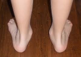
                        The Flexible Flatfoot in the Adults

The adult acquired flatfoot deformity is characterized by flattening of the medial longitudinal arch with insufficiency of the supporting posteromedial soft tissue structures of the ankle and hindfoot.

Aetiology


Although the etiology of this deformity can be arthritic or traumatic in nature, it is most commonly associated with posterior tibial tendon dysfunction (PTTD). Developmental etiologies also may be responsible for a flexible flatfoot deformity. These include conditions associated with soft tissue laxity (Ehlers-Danlos and Marfan syndromes), accessory navicular, and neuro-muscular diseases. Extrinsic factors are less common but can result from trauma involving the medial structures in an eversion type injury.

Two potential mechanical causes of an acquired flatfoot deformity include medial column instability and a contracture of the Achilles tendon or gastrocnemius fascia. With the former, medial column instability results in forefoot varus and a compensatory hindfoot valgus. With the latter, a tight Achilles tendon or gastrocnemius fascia results in transmission of dorsiflexion forces from the ankle to the transverse tarsal joint and midfoot. This leads to midfoot collapse and hindfoot valgus with lateral peritalar subluxation of the navicular and subfibular impingement.

Pathology

An acquired flexible flatfoot deformity is most often associated with Posterior Tibial tendon (PTT) dysfunction. Biomechanic overloading as described above can lead to chronic microtrauma in the tendon. With advancing age, the tendon’s elastic compliance decreases because of changes in collagen structure, thus creating a pathologic sequence where tendon weakening results in failure of the static stabilizers of the arch. Poor blood supply may initiate this process or may prevent an adequate healing response, resulting in chronic inflammation, tenosynovitis, and tendinosis.

Clinical Examination

Patients usually complain of medial ankle and hindfoot pain that radiates to the arch of the foot or proximally to the leg. As the deformity progresses, there may be a complaint of lateral or sinus tarsi pain caused by subfibular impingement. Although some patients will attribute a nonspecific traumatic event to the pain, most patients will relate a gradual onset of the pain with loss of the medial plantar arch over recent months or years.

On physical examination, it is helpful to evaluate the patient in short pants with both shoes off. This allows the clinician to note the alignment of not only the foot and ankle, but also the knee. With genu valgus, an individual’s center of gravity may be altered and more load may be placed on the medial ankle and PTT. Comparison of tread wear on the shoes may reveal more posteromedial wear than the opposite side. On examination of the standing patient from behind, the presence of hindfoot valgus can be noted and measured, and the “too many toes” sign can be identified. The patient should be asked to perform a double leg heel rise so that the presence or absence of hindfoot inversion can be identified. Next, the patient is asked to perform a single leg heel rise on the affected side noting that inability to do so is consistent with PTTD.
Examination sitting should include assessment of ankle and subtalar range of motion. Ankle motion should be measured with the knee extended and flexed with the transverse tarsal joint locked and unlocked. This will allow the examiner to assess for Achilles tendon and gastrocnemius contractures. Palpation of the posteromedial ankle and hindfoot may reveal tenderness, swelling, or fullness. The sinus tarsi, talar dome, and navicular tuberosity should be palpated. Callus formation over the subluxated talar head may be noted. For patients who have a flexible flatfoot, reduction of the talonavicular joint and correction of the hindfoot valgus/forefoot abduction is possible. Lastly, the PTT strength is tested with resistance against the inverted and plantarflexed foot.


Diagnostic Imaging

Clinical examination and radiographs (in weight bearing Position) are usually sufficient to establish the diagnosis of PTTD. In certain instances, however, the use of MRI can be helpful to confirm the diagnosis, evaluate the amount of pathology in the PTT and spring ligament complex, and detect bone edema.


STAGES of PTTD

Stage I consists of painful synovitis of the tendon. Nevertheless, tendon length and function are maintained so there is no deformity.
Stage II disease, there is progressive tendon dysfunction and a flexible flatfoot deformity develops.
Stage III involves a rigid deformity with stiffness and often arthritis of the midfoot and hindfoot.
Stage IV consists of tibiotalar valgus, usually with associated arthritic changes.

Conservative Treatment


The stage and progression of the flatfoot deformity will generally determine the degree and duration of the conservative treatment. The initial treatment of the adult flexible flatfoot deformity (stage II PTTD) focuses on improving symptoms by decreasing the forces transmitted through the posteromedial hindfoot. The patient should be encouraged to lose weight, modify repetitive loading activities, and use supportive shoes.
(Eric Giza, MDa,*, Gerard Cush, MDb, Lew C. Schon, MD)